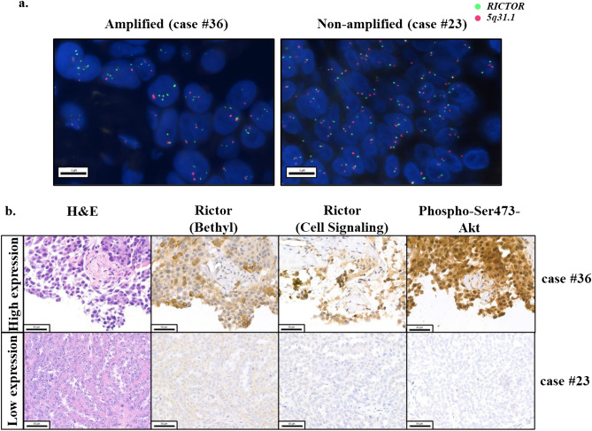
Figure 1.
Representative images of RICTOR-amplified/non-amplified cases by FISH (a) and the high/low expressions of IHC stainings (b) in the studied tumour samples. The presented squamous cell carcinoma of the lung (case #36) showed RICTOR amplification with high Rictor and high Phospho-Ser473-Akt protein expressions. The other tubo-ovarian high-grade serous carcinoma (case #23) represents no amplification without increased Rictor and Phospho-Ser473-Akt expressions.

