Figure 1.
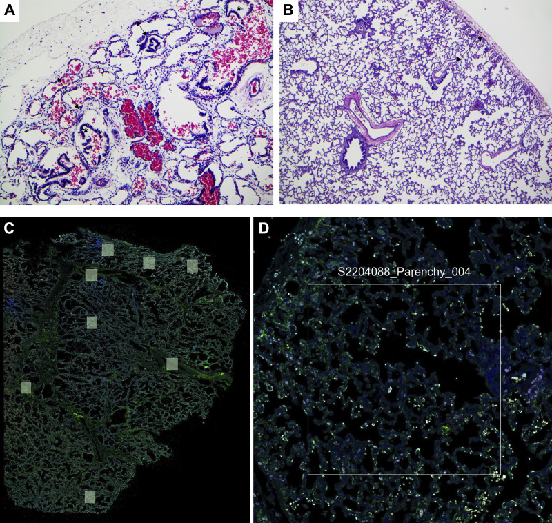
Histopathological characterization of the CDH lungs: representative H&E stained lung tissue sections from CDH (A) and control lungs (B). A: lung section from patient with CDH showing severe pulmonary hypoplasia with airways (*) seen approaching pleural surface and markedly decreased radial alveolar count of 1–2 (double-headed arrows, H&E, ×100). B: lung section from control patient showing appropriately developed pulmonary parenchyma with radial alveolar count of 4–5 (double arrowheads; H&E, ×40). Representative low (C)- and high (D)-magnification lung section stained with a nuclear marker (SYTO 13), PanCK (Pancytokeratin; epithelial marker), and CD45 (immune cell marker) with demarcation of the region of interest for the selection of the distal lung parenchyma. CDH, congenital diaphragmatic hernia; H&E, hematoxylin-eosin.

