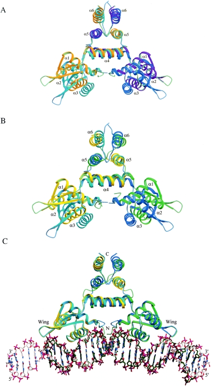
FIG. 3.
Structural comparison. (A) Superposition of unbound BlaI and DNA-bound BlaI dimers. (B) Superposition of unbound BlaI and unbound MecI dimers (1OKR). (C) Superposition of DNA-bound BlaI and DNA-bound MecI (1SAX) complexes. The structures were superimposed as described in the text. Protein molecules are shown as ribbons, and DNA molecules are shown as stick models in which the carbon atoms are in white and green for the DNA bound to BlaI and MecI, respectively. In (A), the unbound BlaI monomers are in cyan and blue, whereas the DNA-bound BlaI monomers are in orange and magenta. In (B) and (C), the two BlaI monomers are in cyan and blue, whereas MecI is in yellow and green.

