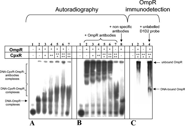
FIG. 4.
CpxR and OmpR bind simultaneously at the promoter region of csgD. Band-shift assays were performed with pure CBP-CpxR, pure OmpR-His6, and a 32P-labeled csgD promoter fragment in the absence (A) or in the presence (B) of antibodies raised to OmpR. D1D2 probe without protein (lanes 1), and D1D2 probe with 1.65 μM OmpR-His6 (lanes 2) are as shown. Increasing amounts of CBP-CpxR were added in the binding reaction containing 1.65 μM concentrations of OmpR-His6. D1D2 probe with 1.65 μM OmpR-His6 and 0.48 μM CBP-CpxR are shown in lanes 3; lanes 4 are the same as in lane 3 but with 0.96 μM CBP-CpxR; lanes 5 are the same as for lanes 3 but with 1.92 μM CBP-CpxR (lanes 5); lanes 6 are the same as for lanes 3 but with 2.88 μM CBP-CpxR; lanes 7 show D1D2 probe with 2.88 μM CBP-CpxR. A control lane contains D1D2 probe with 1.65 μM OmpR-His6 and nonspecific antibodies (panel B, lane 8). (C) Immunodetection of OmpR in DNA complexes after transfer of EMSA gel proteins onto a membrane as described in Materials and Methods. Lanes: 1, D1D2 probe without protein; 2, D1D2 probe with 1.65 μM OmpR-His6 and 1.92 μM CBP-CpxR; 3, D1D2 probe with 1.92 μM CBP-CpxR; 4, D1D2 probe with 1.65 μM OmpR-His6, 1.92 μM CBP-CpxR, and a fourfold excess of unlabeled D1D2 probe.

