Figure 2.
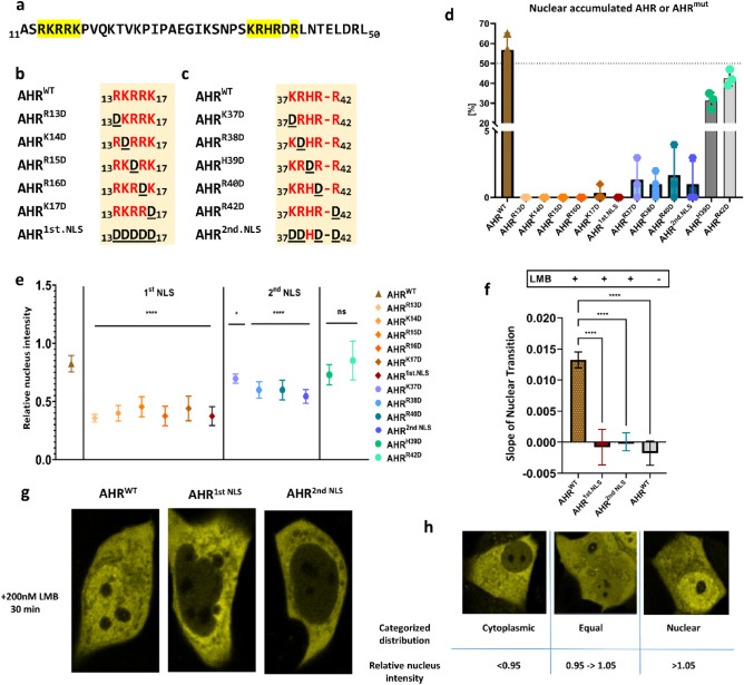
Role of the nuclear localization signal (NLS) on the basal shuttling of AHR. The N-terminal bHLH domain of AHR is indicated as amino acid sequence (a). Graphical representation of mutated amino acids in 1st NLS (b) and 2nd NLS (c). The percentage of cells with nuclear accumulated AHRWT or AHRmut for 100 randomly chosen cells in the basal state (d) according to our classification (h). The ratio of the mean fluorescence intensity within the nucleus and the mean fluorescence intensity of the cells in the cytoplasmic state is given as relative nuclear intensity (e). Slopes of the nuclear transition of AHRWT or AHRmut after treatment with 200 nM leptomycin B (LMB) for 30 min (f). Values represent the mean ± S.D of n = 100 (d), n = 10 (e), n = 14 for AHRWT and n = 5 for AHRmut or unstimulated AHRWT (f). Statistical analysis was performed with a one-way ANOVA, Dunnett's post-test, *p < 0.05, ****p < 0.0001.

