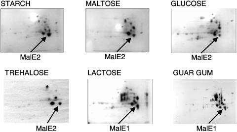
FIG. 3.
Results of 2-D gel electrophoresis of periplasmic extracts derived from cells grown on lactose, guar gum, starch, maltose, trehalose, and glucose. The spots corresponding to either MalE1 or MalE2 are indicated. The spot from trehalose-grown cells showed mass peaks corresponding to both proteins. Proteins were separated based upon molecular mass (vertically) and isoelectric point (horizontally).

