
Figure 3: Binding of PTH(1–34) to the extracellular domain (ECD) of PTH1R.
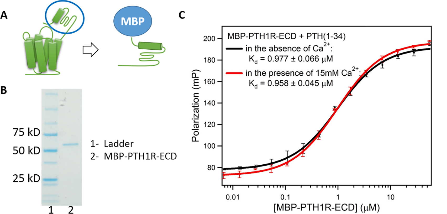
(A) Construction of MBP-PTH1R-ECD. (B) SDS-PAGE gel of MBP-PTH1R-ECD shows Lane 1, markers; lane 2, purified MBP-PTH1R-ECD (62 kD) (C) Binding of PTH(1–34) to MBP-PTH1R-ECD measured by fluorescence polarization. (Buffer: 50 mM Tris-HCl, pH 7.5, 150 mM NaCl, 1 mM EDTA). Each curve shows the average of three fluorescence polarization experiments.