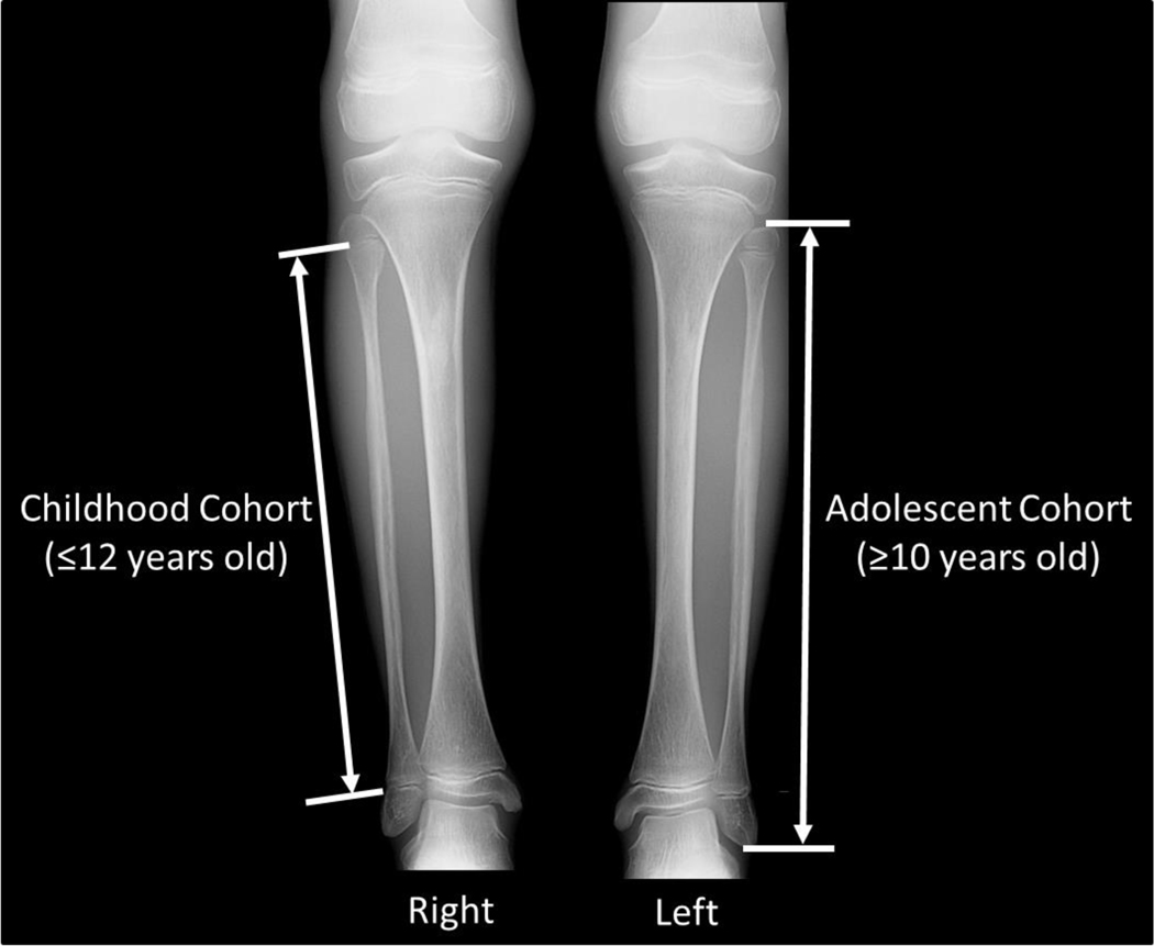
Fig. 1.
Methodology for long bone length measurements, using lower extremity radiographs of a 10-year 11-month-old girl with Progeria as illustration. For the childhood cohort (≤12 years old), bone length measurements were made between the physes along the midline long axis of the bone (right fibula measurement of above). For the adolescent cohort (≥10 years old), bone length measurements were made along the midline long axis of the bone from the upper margins of the proximal to the lower margin of the distal ossified epiphyses (left fibula measurement of above). This allowed for overlap in the 10–12 year old range.

