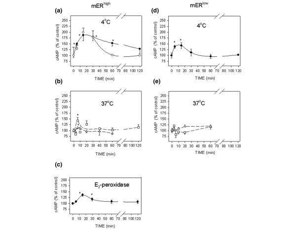
Figure 2.

Optimization of conditions for 17β-estradiol (E2)-induced cAMP accumulation and measurement. (a–c) MCF-7 cells enriched for membrane estrogen receptor-α (mERhigh) and (d, e) MCF-7 cells depleted for membrane estrogen receptor-α (mERlow). All of the cells were stimulated with 1 pmol/l E2, or an equivalent amount of E2 conjugated to peroxidase, for different time intervals, and the intracellular cAMP levels were assessed. (Panels a and d) Cells were incubated at 4°C in defined medium (DM) either attached to a plate (open circles) or in suspension (closed circles). (Panels b and e) Attached cells were incubated at 37°C in DM medium (triangles) or DCSS medium (medium with 4 × dextran-coated charcoal-stripped serum; squares). (Panel c) Cells in suspension were stimulated with E2-peroxidase at 4°C. All experiments were repeated at least three times, and each time point was in triplicate. The data are presented as means ± standard error and the asterisks represent significant differences (P < 0.05) as compared with time 0.
