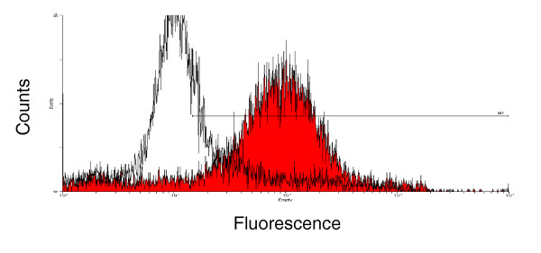
Figure 1.

Flow cytometric analysis of A2L2 cells with serum from mice vaccinated with virus-like replicon particles (VRP)-neu or VRP-hemagglutinin (HA). Serum was collected 2 weeks after a single vaccination of Balb/c mice with 106 IU VRP-neu (filled curve) or 106 IU VRP-HA (open curve). The primary serum was diluted with PBS (1:100), and FITC-labeled goat anti-mouse IgG diluted in PBS (1:1000) was used as the secondary antibody.
