Figure 10.
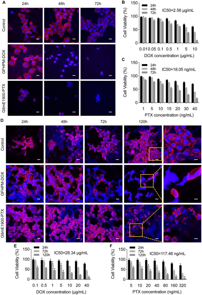
Pharmacodynamic evaluation of drug delivery nanocarriers on scaffold-derived 3D lung cancer models. (A) Morphology of 2D cells treated with GFHPM-DOX and G8mE1900-PTX micelles at 24, 48 and 72 h, respectively. Scale bar 20 μm. (B) Cell viability of 2D cells treated with GFHPM-DOX micelles (n = 3). (C) Cell viability of 2D cells treated with G8mE1900-PTX micelles (n = 3). (D) Morphology of 3D spheroids treated with GFHPM-DOX and G8mE1900-PTX micelles at 24, 48, 72 and 120 h, respectively. Scale bars 20 μm for low-magnification field and 10 μm for high-magnification field. (E) Cell viability of 3D spheroids treated with GFHPM-DOX micelles (n = 3). (F) Cell viability of 3D spheroids treated with G8mE1900-PTX micelles (n = 3). The results showed that the 3D lung cancer model is more tolerant to antitumor drugs than the conventional 2D model cells.

