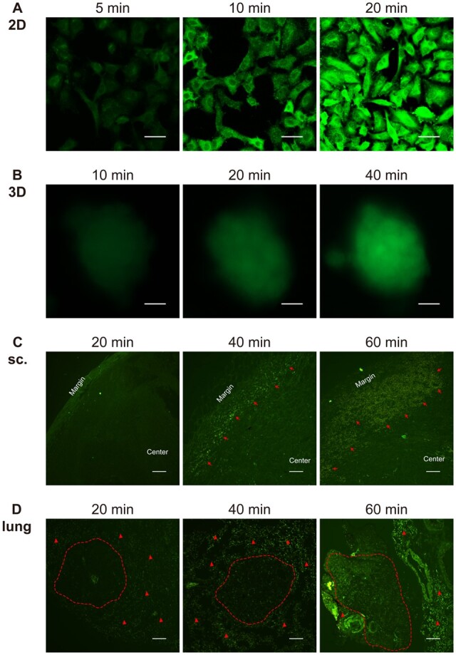
Figure 8.

The penetration and distribution of positively charged nanocarriers in 2D-cultured cells (A, scale bar 20 μm), 3D spheroids (B, scale bar 200 μm), subcutaneous xenograft tumors (sc.) (C, internalization of G8mE1900-FITC micelles are indicated by red arrows, scale bar 200 μm), and in situ lung cancer models (D, nodules are circled by the red dotted line, red triangles indicate the accumulation and distribution of G8mE1900-FITC micelles in the lung interstitial, scale bar 200 μm). the results showed that positive nanocarriers penetrated and distributed faster in 2D-cultured cells than in 3D spheroids and in vivo models, with 3D spheroids showing a similar pattern to the in vivo models.
