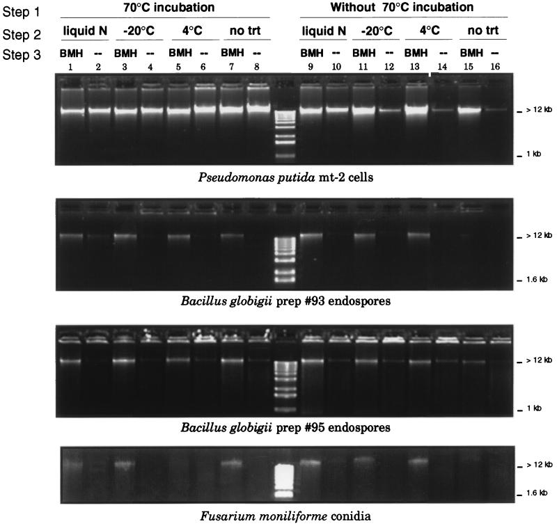
FIG. 1.
Relative amounts of DNA extracted from P. putida cells, B. globigii endospores, and F. moniliforme conidia processed by variations of the three-step procedure described in Materials and Methods. (Step 1) Cells or spores were suspended in TENS buffer and either incubated at 70°C for 20 min, or this step was omitted and samples were immediately carried to step 2. (Step 2). Samples were frozen in liquid nitrogen to −20°C or chilled to 4°C and then rapidly heated in a boiling water bath. This step was repeated three times or was not done (no trt). (Step 3) Samples were either homogenized for 3 min at 5,000 rpm in a mini-bead beater (BMH), or this step was omitted (--). Equal volumes of DNA extract were separated by agarose gel electrophoresis on a 3% agarose gel, and DNA was visualized under UV light after staining with ethidium bromide. Comparison of the DNA band intensities in a gel illustrates the different amounts of DNA released from equal amounts of starting material by each procedure or procedure combination. Lane 16 in each panel illustrates the combined amount of DNA present as extracellular background DNA and released by room temperature incubation in TENS buffer containing the detergent SDS. prep, preparation.

