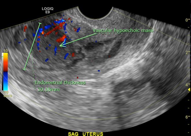
FIGURE 1.

Transvaginal gray‐scale ultrasound image of the uterus in the sagittal plane. The endometrial stripe is markedly thickened to 30 mm with an irregular contour delineated by a small amount of fluid within the endometrial cavity. There is a 3.2 × 2.2 × 3.3 cm3 ovoid homogeneous hypoechoic mass within the anterior endometrium with internal color Doppler flow and a low resistance arterial waveform.
