FIGURE 2.

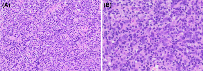
Photomicrograph of the endometrial biopsy. Sheets of large lymphoma cells are present, with atrophic background endometrium. [H&E stain, original magnification, ×200 (A), ×400 (B)].

Photomicrograph of the endometrial biopsy. Sheets of large lymphoma cells are present, with atrophic background endometrium. [H&E stain, original magnification, ×200 (A), ×400 (B)].