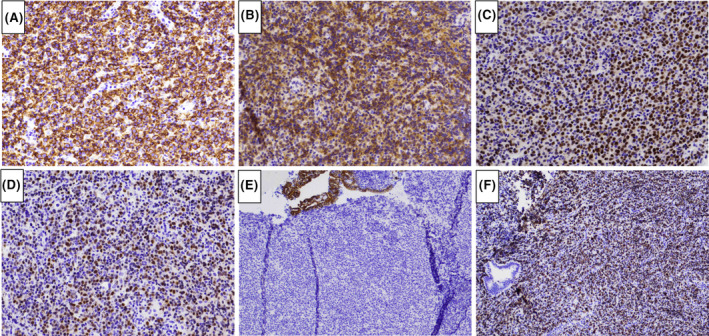
FIGURE 3.

Immunohistochemical characterization of the lymphoma. The lymphoma cells are positive for CD20 (A), CD10 (B), BCL6 (C), and MUM1 (D), and negative for cytokeratin CAM5.2 (E). Ki67 stain showed a high proliferation rate (about 70% of lymphoma cells in cycle) (F). [Immunoperoxidase staining; original magnification, ×400 (A–D), ×100 (E, F)].
