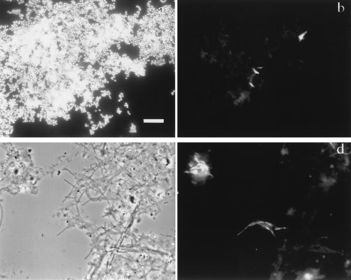
FIG. 5.
Phase-contrast and epifluorescence images after FISH of mixed cultures and activated sludge. Bar = 10 μm. (a and b) Mixture of cultures of R. rhodochrous and G. amarae SE-102 hybridized with TRITC-labeled probe S-S-G.am-0205-a-A-19 and dual stained with DAPI. Panels a and b show the same image field, as viewed by epifluorescence microscopy, prepared with the DAPI and TRITC filter sets, respectively. (c and d) RAS sample from San Francisco Southeast Water Pollution Control Plant hybridized with probe S-S-G.am-0205-a-A-19 labeled with TRITC. Panels c and d show the same image field, as viewed by phase-contrast microscopy and epifluorescence microscopy, respectively.

