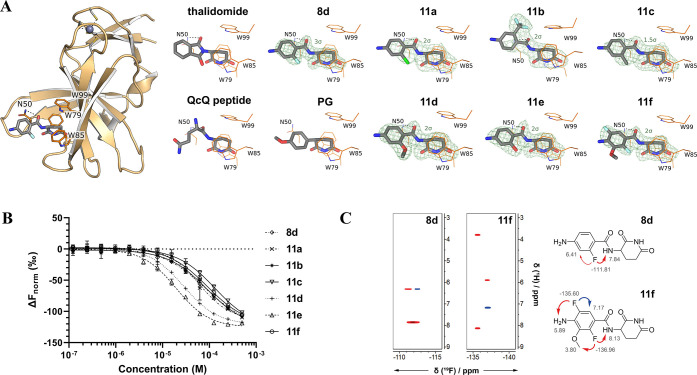
Figure 2.
(A) Difference electron density (Fo – Fc) maps, contoured at the indicated sigma level, of ortho-substituted benzamide compounds bound to crystallized MSCI4. The full-size crystal structure is exemplary shown for 8d on the left. For comparison, thalidomide, a QcQ peptide (the two last amino acids of a natural degron peptide), and a phenyl glutarimide (PG) are shown (PDB identifiers: 4V2Y, 8BC7, and 7SHH, respectively). Nitrogen is shown in blue, carbon in gray, oxygen in red, fluorine in light blue, and chlorine in chartreuse. (B) Dose–response curves for compounds 8d and 11a–11f were obtained in competitive MST measurements with BODIPY-uracil and hTBD (n = 3). (C) Sections of 19F,1H-HOESY NMR spectra of 8d and 11f, respectively. Spatial proximity between F and the amide NH provides evidence for intramolecular interactions. NOE with residual water is not displayed.

