
Fig. 3.
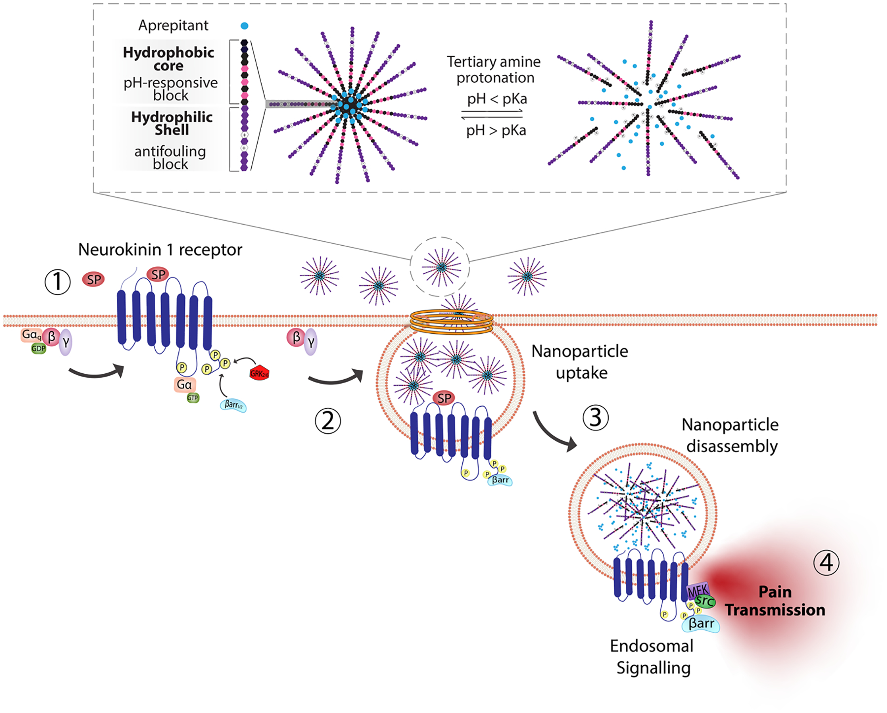
pH-responsive nanoparticles for endosomal targeting of the NK1R. pH-responsive nanoparticles were designed using amphiphilic diblock copolymers formed by four monomers. The hydrophobic portion of the diblock copolymer is formed by di(ethylene glycol) methyl ether methacrylate (DEGMA) and the pH-responsive monomer 2-(diethylamino)ethyl methacrylate (DIPMA). The hydrophilic portion is formed by poly(ethylene glycol) monomethyl ether methacrylate (PEGMA) and a positively charged monomer, 2-(dimethylamino)ethyl methacrylate (DEAEMA). The antagonist aprepitant was physically entrapped in the core of the nanoparticles. (1) The NK1R is activated by substance p (SP) and (2) couples to G-protein, followed by (3) rapid endocytosis. (4) From this location, NK1R transmits pain. Nanoparticles are passively endocytosed, and the acidity of endosomes triggers the disassembly of nanoparticles and release of aprepitant from the core. Released aprepitant can now antagonize the endosomal signal produced by the NK1R to decrease pain transmission.