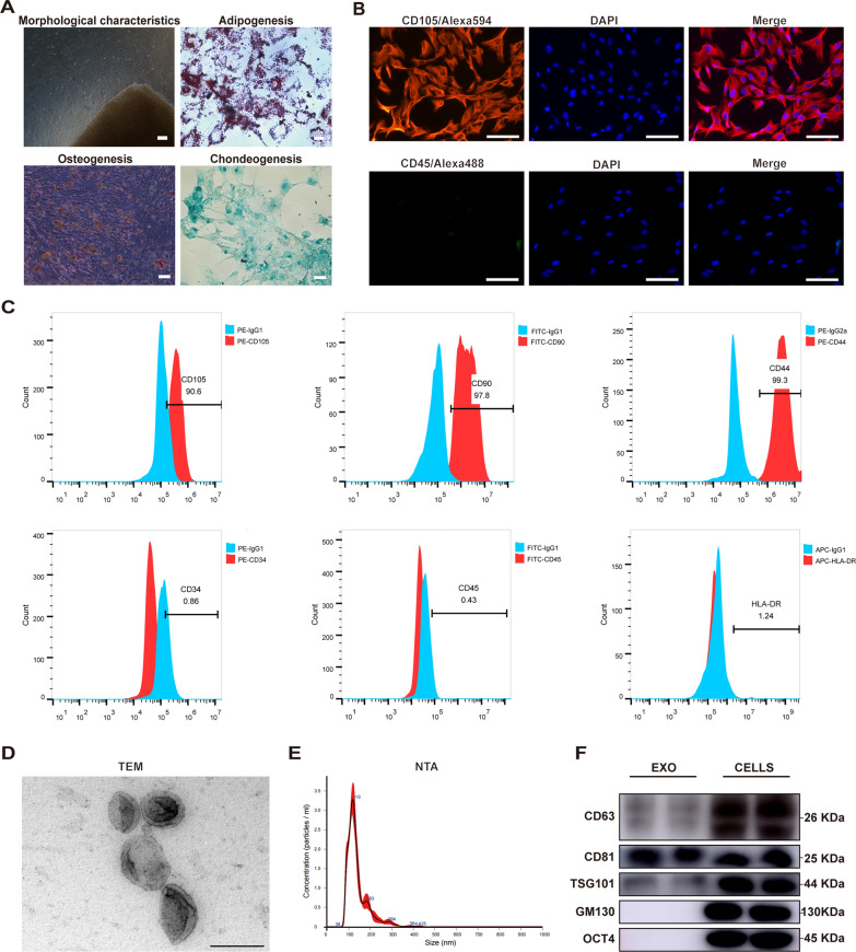
Fig. 1.
Isolation and characterization of human umbilical cord mesenchymal stem cells (hucMSCs) and hucMSC-exos. A The morphology of hucMSCs isolated and cultured by tissue block attachment at the third passage (100x), the Oil Red O staining of adiopogenic identification (400x), the Alizarin Red staining of osteogenic identification (100x) and the Alcian blue staining of chondrogenic differentiation (200x). B Immunofluorescence (IF) staining of CD105 and CD45 of hucMSCs. Scale bars, 50 μm. C Positive expression of CD105, CD90, and CD44 and negative expression of CD34, CD44, and HLA-DR were detected by flow cytometry. The blue peak represented the isotype control antibody and the red one represented the MSC-associated markers. D Representative transmission electron microscopy (TEM) image of hucMSC-exos. Scale bar, 200 nm. E The particle size and concentration of hucMSC-exos measured by nanoparticle tracking analysis (NTA). F Representative western blot images of exosomal-associated (CD63, CD81, TSG101), Golgi-associated (GM130) and MSC-associated (OCT4) markers in hucMSC-exos and hucMSCs

