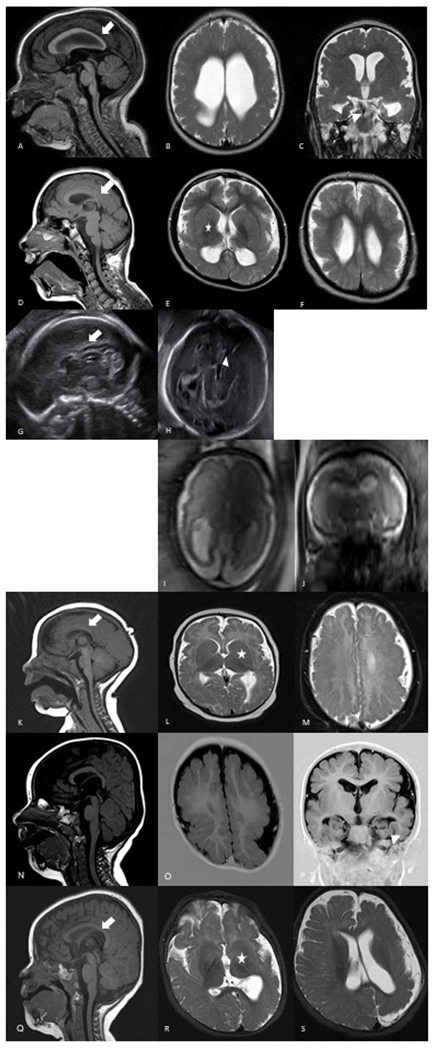
Figure 1.

Brain imaging findings of individuals with heterozygous pathogenic variants in GRIN1 and GRIN2B. Individual 1 has diffuse dysgyria, hypoplastic corpus callosum (arrow) and dysmorphic hippocampi (arrowhead) (A–C). Individual 2 has bilateral dysgyria, hypoplastic corpus callosum (arrow) and dysmorphic basal ganglia (asterisk) (D–F). Prenatal ultrasound in individual 3 at 25 gestational weeks shows absent corpus callosum (arrow) and dilated third ventricle (arrowhead) (G–H). Individual 4 has diffuse dysgyria and enlarged lateral ventricles on prenatal MRI at 29 gestational weeks (I–J). Individual 5 presents with bilateral dysgyria, enlarged basal ganglia (asterisk) and hypoplastic corpus callosum (arrow) (K–M). Individual 6 has bilateral symmetric dysgyria and dysmorphic hippocampi (arrowhead) (N–P). Individual 7 has asymmetric dysgyria of the left hemisphere, dysmorphic basal ganglia (asterisk) and corpus callosum (arrow) (Q–S).
