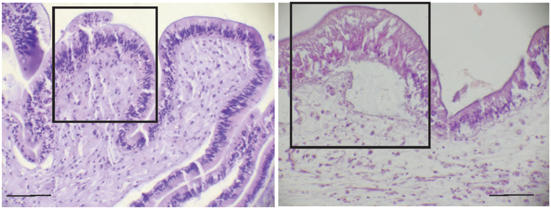
Figure 5.

Common lesions noted in the epidermis and dermis. On the left (Pisaster ochraceus), within the box, there is multifocal epidermal degeneration and lytic necrosis characterized by a loss of cell distinction, nuclear condensation, and fragmentation (karyorrhexis). On the right (Pycnopodia helianthoides), there is coagulation-type epidermal necrosis characterized by loss of cellular detail, particularly nuclear and cell margin distinction with increased uptake of eosin stain (pink staining) by the cytoplasm. Both types of lesions were associated with microscopic epidermal loss or ulceration in some specimens. The right image also shows the two most common lesions within the dermis: separation of the connective tissue fibers by edema and infiltrations of coelomocytes consistent with inflammation. A low instance of dermal degeneration and necrosis also occurred. Scale bars = 50 μm.
