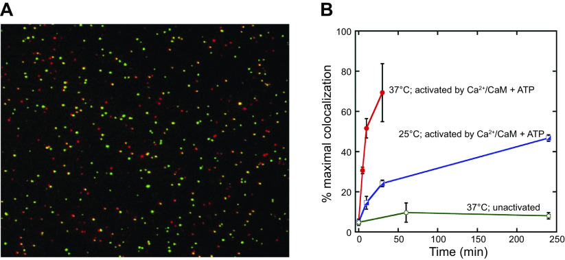
Figure 11.
Single-molecule assay for subunit exchange reveals activation-dependent subunit exchange. A: a representative single-molecule total internal reflection fluorescence (TIRF) image, with red and green channels overlaid. B: the rate of increase in colocalization is significantly faster at 37°C (red) compared to 25°C (blue) when Ca2+/calmodulin (CaM) and ATP are added. At 37°C, the unactivated sample (i.e., with no addition of Ca2+/CaM and ATP) shows only a low level of exchange even at long time points (green). Image from Ref. 406, with permission from eLife.

