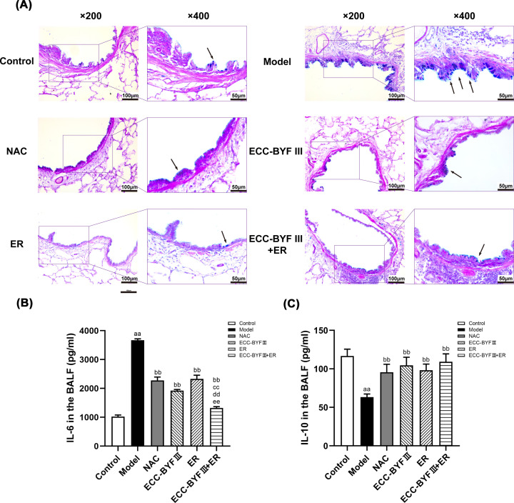
Figure 4. Changes of goblet cells in airway and changes of IL-6 and IL-10 in all groups.
(A) AB-PAS staining photos of the airway in all groups (AB-PAS; ×200, ×400).→: goblet cells. (B) Changes of IL-6 in all groups. The data are expressed as the mean ± SE, (n=6). aaP<0.01 vs. the normal control group; bbP<0.01 vs. the disease model group; ccP<0.01 vs. the NAC group; ddP<0.01 vs. the ECC-BYF III group; eeP<0.01 vs. the ER group. (C) Changes of IL-10 in all groups. The data are expressed as the mean ± SE (n=6). aaP<0.01 vs. the normal control group; bbP<0.01 vs. the disease model group.

