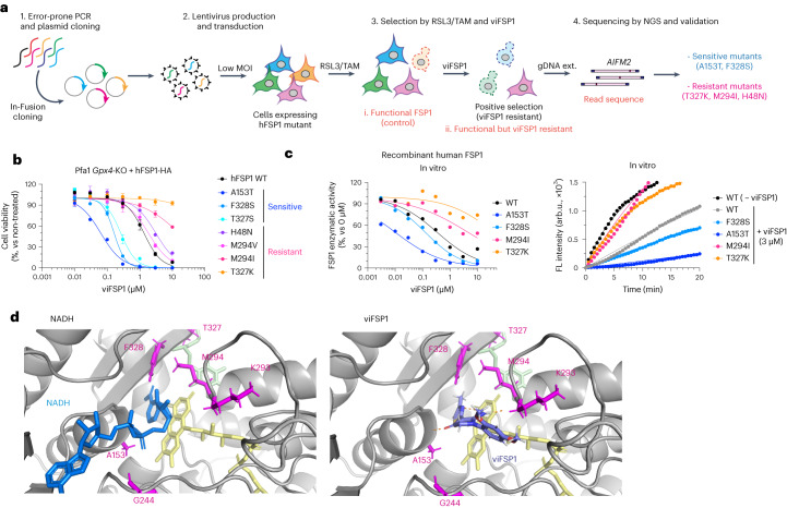
Fig. 5. viFSP1 targets the NAD(P)H-binding pocket of FSP1.
a, Schematic of the mutational screen to identify the responsible sites affecting viFSP1 activity in hFSP1. b, Viability in Pfa1 Gpx4-KO cells stably overexpressing mutant hFSP1-HA, treated with viFSP1 for 24 h. Data are shown as the mean ± s.d. of 3 wells of a 96-well plate from 1 of 3 independent experiments (See also Supplementary Videos 1–4). c, Representative dose–response curves for the effect of viFSP1 on the activity of WT FSP1 or its mutants, using recombinant purified hFSP1 protein (left). Representative in vitro assays of inhibition of WT FSP1 and the mutant variants by 3 µM viFSP1 or 0 µM viFSP1 (right). Data are from a single well of a 96-well plate from 1 of 3 independent experiments. d, Comparison between viFSP1 and NADH in the respective binding pockets. A153, G244, M294, T327 and F328 are highlighted in magenta. In silico simulation identified the interaction between the main chains of A153, K293 and viFSP1 using a hydrogen bond. Each interaction is depicted by the dashed line in orange.

