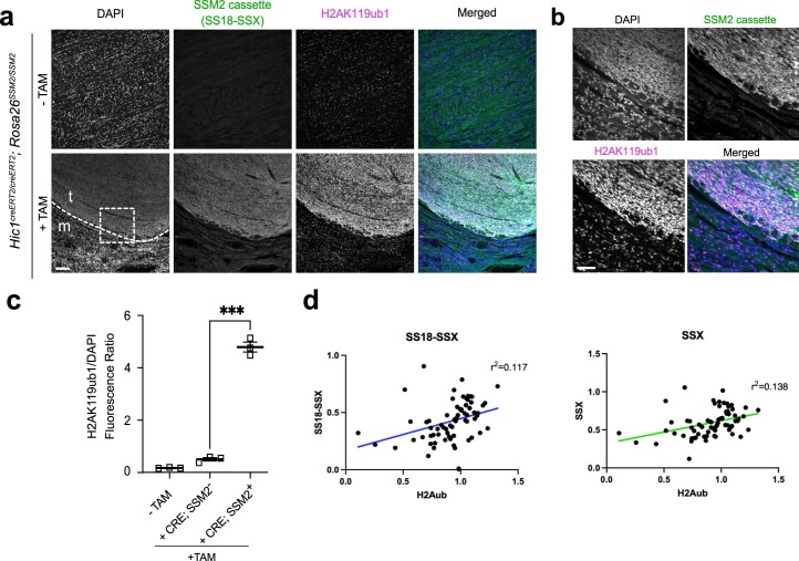
Extended Data Fig. 5. Murine and human synovial sarcomas exibit high levels of H2AK119ub1.
a) Immunofluorescence of Hic1creERT2/creERT2; Rosa26SSM2/SSM2 mice at 16-week endpoint tongue tissue showing left, non-tamoxifen treated mice (-TAM) (upper panel) or tamoxifen treated mice expressing or not the SSM2 cassette (human SS18-SSX2) embedded in striated muscle +TAM; SSM2+ and +TAM; SSM2− cells (lower panel). The cells are stained for DAPI, SSM2 and H2AK119ub1. The scale bar represents 100 um. b) Close-ups of images shown in the panel above, in the area delineated by the dashed square in (a). c) Quantification of H2AK119ub signal intensity normalised to DAPI signal intensity in 3 biological replicates (3 different mice) in non-tamoxifen treated mice (-TAM), or tamoxifen treated mice ( + TAM) expressing or not the SSM2 cassette (human SS18-SSX2) and showing normal tongue muscle ( + TAM; SSM2−) adjacent to synovial sarcoma tumours ( + TAM; SSM2+). Asterisks represent p-values of paired one-tailed t-test between groups (p = 0.0006). d) Spearman correlation between SS18-SSX, left or SSX, right signals and H2AK119ub1 signals per sarcoma sample.

