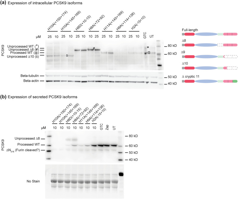
Figure 2.
PCSK9 protein isoform expression was assessed in Huh-7 cells after treating with PMOs for 3 days. Western blot analysis of (a) intracellular PCSK9 protein isoforms and (b) secreted PCSK9 isoforms after targeted exon skipping. Dated replicate experiments are shown in Supplementary Fig. 6. The schematics of the predicted PCSK9 isoforms after exon skipping are shown on the right. The domain(s) predicted to be absent are shown in grey dotted lines. The location of new amino acids introduced into PCSK9 are shown in dark green. WT; wild-type, GTC; sample treated with Gene Tools control PMO, UT1; sample underwent Neon electroporation without any PMO, UT2; untreated sample. The images were cropped for presentation. Full-length original images are shown in Supplementary Fig. 3.

