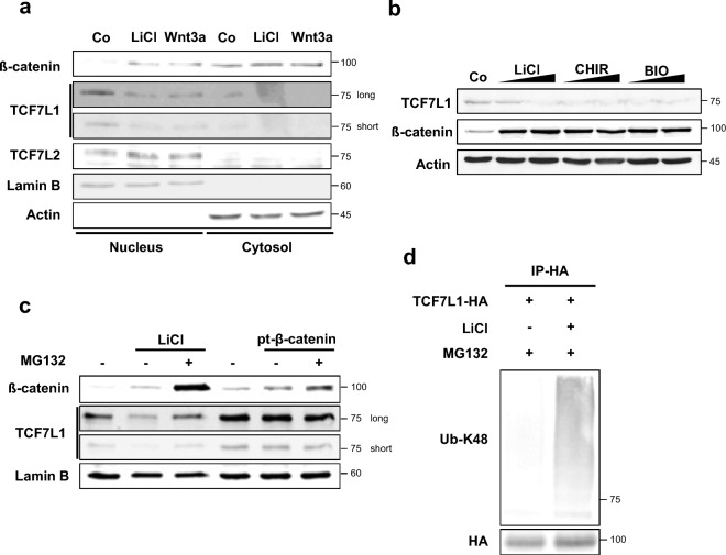
Figure 1.
TCF7L1 undergoes degradation through ubiquitination following activation of the Wnt signaling pathway. (a) Western blot analysis was performed using HeLa cells. Cells were treated with 50 mM LiCl or mWnt3a conditioned media for 16 h. The cytosolic fraction was lysed with CB buffer, while the nuclear fraction was lysed with RIPA buffer following initial lysis with CB buffer. (b) HeLa cells treated with LiCl (25 mM and 50 mM), CHIR99021 (5 μM and 10 μM) and BIO (5 μM and 10 μM) conditioned media for 16 h and lysed with RIPA buffer. (c) Western blot analysis was performed using HeLa cells. Cells were treated with LiCl-conditioned media or transfected with the pt-β-catenin plasmid, followed by treatment with 10 μM MG132 for 12 h prior to harvesting. (d) In vitro ubiquitination assay was performed using control HeLa cells. TCF7L1-HA (6 μg) was transfected into HeLa cells. Then, the cells were treated with 50 mM LiCl-conditioned media for 16 h, followed by treatment with 10 μM MG132 for 12 h prior to harvesting with IP buffer. The cells were lysed with the IP buffer and subjected to immunoprecipitation with an HA antibody. Ubiquitination levels were determined by anti-UB-K48.

