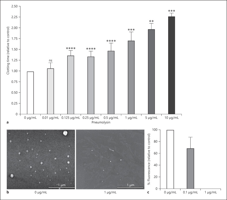
Fig. 7.
PBMC-derived procoagulant MVs after incubation with pneumolysin. PBMCs were stimulated with LPS for 24 h, and MVs were purified from the supernatant as described in methods. a MVs were incubated with indicated concentrations of pneumolysin or buffer (0 μg/mL) for 20 min at 37°C. MVs were then added to re-calcified plasma, and the time for clot formation was determined. Clotting times were normalized for each donor in relation to the control (0 μg/mL), which value was set to 1. b SEM of procoagulant MVs treated with buffer (0 μg/mL) or 1 μg/mL pneumolysin (scale bar, 1 μm). c MVs were stained with PKH26 and incubated with indicated pneumolysin concentrations for 20 min at 37°C. Fluorescence was detected in SpectraMax. Absorbance was normalized for each donor in relation to the MV control (0 μg/mL), which value was set 100%. Values represent the mean ± SD of MVs from four different donors. Significance values calculated in reference to the control using the one-way ANOVA with Dunnet's posttest test **p < 0.005, ***p < 0.0002, ****p < 0.0001. SEM, scanning electron microscopy.

