Fig. 1.
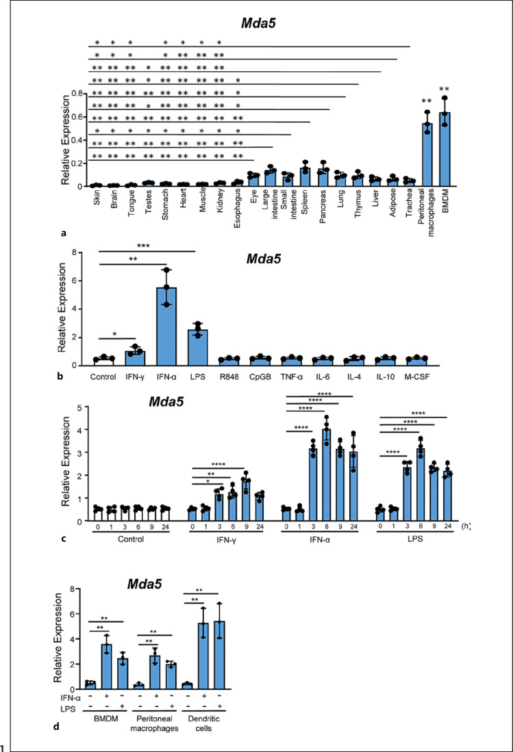
Selective tissue expression of Mda5. a Tissues from three mice were used to obtain RNA, and Mda5 expression was determined by RT-PCR (independent experiments, n = 3). For comparison, we used BMDMs and peritoneal macrophages that were significantly increased in relation to all the tissues (p < 0.01). b Seven-day-old BMDMs were starved of growth factors for 16–18 h and then incubated for 24 h with different stimuli at a concentration of 10 ng/mL with the exception of R848 and CpG which was 2.5 μg/mL. Mda5 expression was then determined by RT-PCR (n = 3). Controls were untreated cells. c Time course of Mda5 expression. BMDMs were incubated with the indicated stimuli, and Mda5 induction was measured by RT-PCR (n = 4). d Mda5 expression is induced in BMDM, peritoneal macrophages and DCs by IFN-α or LPS. Cells were incubated for 24 h with the indicated stimuli and Mda5 expression was then determined by RT-PCR (n = 3). Each experiment was performed in triplicate, and the results are shown as the mean ± SD. *p < 0.05, **p < 0.01, ***p < 0.001, and ****p < 0.0001 in relation to each stimulated sample compared to controls in each experiment. Data were analyzed using the unpaired Student's t test with the exception of c that was calculated using a two-way ANOVA test followed by a Bonferroni correction.

