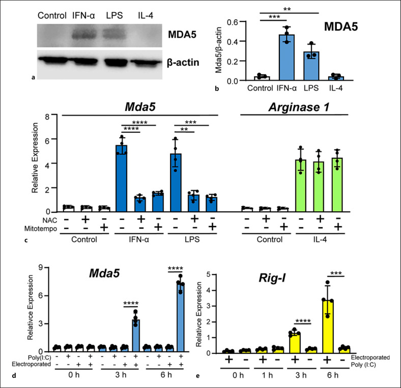
Fig. 2.
Protein and mRNA expression of MDA5. a Representative blot of MDA5 protein expression in BMDMs. Total protein extracts from BMDMs treated for 24 h with the indicated stimuli were subjected to western blotting to determine MDA5 expression. β-actin was used as the loading control. b Quantitation of MDA5 expression in three independent experiments in relation to β-actin expression (n = 3). c BMDMs were incubated for 1 h with media, NAC (20 mM) or Mito-TEMPO (50 μM). The BMDMs were then treated with IFN-α or LPS for 3h and Mda5 expression was determined (n = 4). As control, BMDMs were treated with IL-4 and Arginase 1 was determined (n = 4). d BMDMs were electroporated with poly(I:C) (2 μg in 100 μL), while controls were non-electroporated BMDMs. The mock transfection controls were the electroporated cells with media. Mda5 expression was determined at the indicated times (n = 4). e Similar experiment to that in (d), but Rig-I expression was determined instead (n = 4). Each experiment was performed in triplicate, and the results are shown as the mean ± SD. **p < 0.01, ***p < 0.001, and *p < 0.0001 in relation to each stimulated sample compared to controls in each experiment. Data were analyzed using the unpaired Student's t test.

