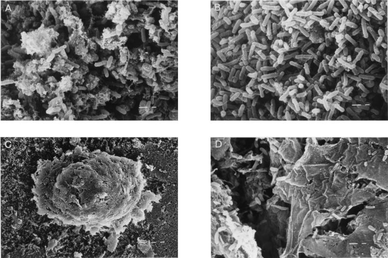
FIG. 1.
SEM micrographs of the midgut region and feces of F. candida, fed with sterile substrate. Microbial colonization of the food bolus (A) and peritrophic membrane (B), as detected in the midgut, is shown. The fecal pellet (C) with excreted fragments of the peritrophic membrane and colonized with bacterial cells (D) is also shown. Left bars in panels A, B, and D, 1 μm; left bar in panel C, 10 μm.

