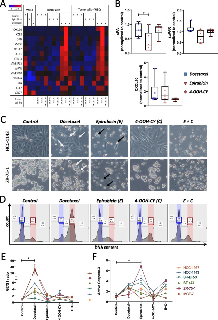
Fig. 3.
Effect of docetaxel, epirubicin, and cyclophosphamide on in vitro cultures of breast cancer cells. A Heat map showing the effect of epirubicin and docetaxel on the release of soluble immune mediators. Epirubicin, docetaxel, or medium as negative control were added to three different breast cancer cell lines (SK-BR3, MCF7, and HCC1143) which were then cultured for 48 h then white blood cells (WBC) were added and the co-culture was cultivated for additional 24 h. The concentration of 14 different immune mediators in the supernatant was determined using a bead array immunoassay. The red colored dots indicate high concentrations of the respective mediator. B Release of uPA, suPAR, and CXCL10 from HCC-1937, HCC-1143, SK-BR-3, BT-474, ZR-75–1, and MCF-7 cells in response to docetaxel, epirubicin, and 4-OOH-CY. C Phase contrast microscopic images of HCC1143 and ZR-75–1 cells after a 48 h cultivation in the presence of docetaxel, epirubicin, 4-OOH-CY, or a combination of the latter two. The white arrows indicate multinucleated giant cells. The black arrows point to apoptotic cells surrounded by apoptotic bodies. D Cell cycle analysis: Cells were treated as described in C and the intracellular DNA was stained with PI. The graph shows the distribution of the fluorescence intensities as assessed by flow cytometry. G1, G2, and S indicate the respective phase of the cell cycle. E Summary of the data shown in D. The results are expressed as mean G2/G1 ratio of the respective cell line (± SD, n = 3). F Quantification of apoptosis. Cells were treated as described in C, then the membrane was permeabilized and stained with antibodies against active caspase 3. The fluorescence was analyzed by flow cytometry. The graph indicates the mean relative distribution normalized to the untreated control (± SD, n = 3). The significance was calculated using a one-way ANOWA overall cell lines

