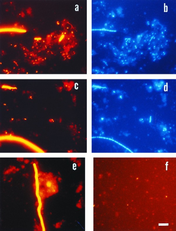
FIG. 2.
Epifluorescence micrographs of bacteria in sediment samples from the Jadebusen Bay of the German Wadden Sea. (a) Hybridization with probe EUB338, specific for Bacteria. (b) Same microscopic field as in panel a with UV excitation (DAPI staining). (c and d) Identical microscopic fields with probe SRB385 (c) and DAPI staining (d). (e) Hybridization with probe DNMA657, specific for Desulfonema. (f) Specific hybridization for Arcobacter with probe ARC94. Bar, 10 μm (applies to all panels).

