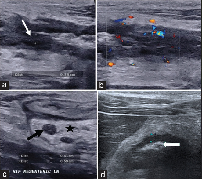
Figure 1.

Intestinal ultrasound images show (a) thickened bowel wall with loss of bowel wall stratification (white arrow) with (b) increased bowel wall vascularity, (c) enlarged lymph node (black arrow) and increased mesenteric echogenicity indicating mesenteric fibrofatty proliferation (star) as well as (d) narrowing or stenosis of the bowel lumen (white arrow) with mild thickened bowel
