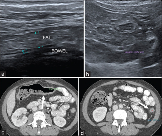
Figure 2.

False negative case example of a 43-year-old with history of Ulcerative Colitis (UC) shows (a) increased fat thickness but no mesenteric echogenicity with normal bowel wall thickness that measures 3 mm at transverse colon. (b) IUS images at descending colon shows mild thickened bowel (at upper limit of normal thickness) with no loss of stratification; thus, concluded as no active disease on ultrasound (c) Axial CT abdomen taken a day after shows mild thickened bowel wall of the transverse colon as well as collapsed and mildly thickened descending colon (white arrows in d). There is mild mesenteric fat streakiness. This patient had clinically active disease of UC at the time of imaging
