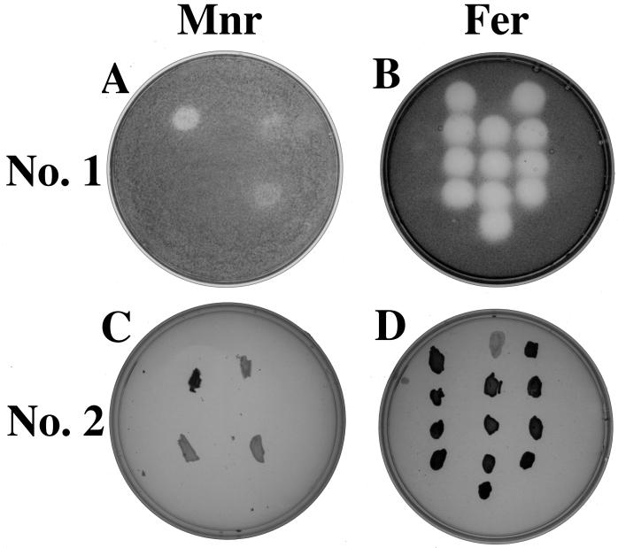
FIG. 3.
(A and C) Plate images of Mnr mutants after application of Mnr screening techniques 1 (anaerobic) (A) and 2 (aerobic) (C) with strains oriented as follows: S. putrefaciens wild type, upper left; anaerobic respiratory mutant T121, upper right; Mnr mutant 48-4, bottom left; Mnr mutant 10-10, bottom right. (B and D) Plate images of Fer mutants after application of Mnr screening techniques 1 (anaerobic) (B) and 2 (aerobic) (D) with strains oriented as follows (from top to bottom): wild type, B29, B41, and A5 (lefthand column); T121, B31, B43, A27, and C23 (middle column); B25, B39, B45, and B101 (righthand column). Note that black color intensity corresponds to benzidine blue color intensity with each screening technique.

