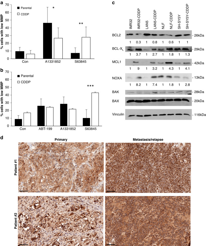
Fig. 3. Shifted response to BH3-mimetics is associated with altered apoptotic signalling.
IMR-32 (a) or Lan5 (b) cells were treated with ABT199, A1331852 or S63845 (1 μM) for 24 h before analysis of MMP using staining with TMRM and flow cytometry. Data shown are mean + SD (n = 4). Statistical analysis was done using t-test (*p < 0.05, **p < 0.01, ***p < 0.001). c Protein expression of parental and CDDP-resistant cells was analysed by Western blotting. One representative blot out of three independent experiments is shown. All experiments were quantified by densitometry and the average protein expression is indicated below each blot. d Neuroblastoma tumour tissues obtained from two individual patients at initial diagnosis (primary) or relapse (metastasis/relapse) was stained with anti-MCL1 antibody. Brown colour indicates positive MCL1 staining. Scale bar represents 100 μm.

