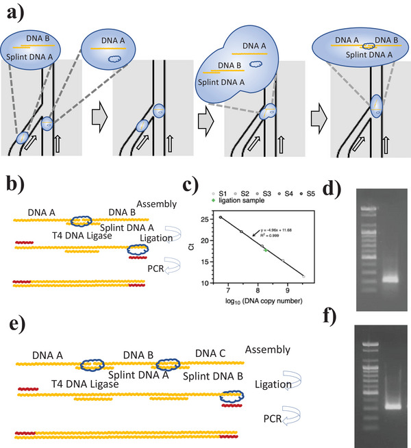
Figure 3.

DNA droplet assembly using splint ligation. a) The DNA droplet in the DCF system. b) The splint ligation process with DNA A and B. c) Standard qPCR curve for the 200‐bps ligation sample (aliquoted to 1/40 000 for the droplet, n = 3). d) The gel electrophoresis results of the splint DNA‐ligated 200 bps fragment. e) The two series splint ligation experiments (DNA A, B, and C), and f) The gel electrophoresis results of the splint DNA‐ligated 300 bps fragment.
