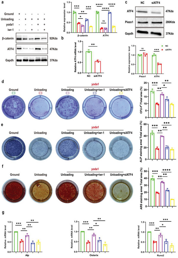
Figure 6.

The critical role of Piezo1/β‐catenin in the function of Gli1+ BMSCs is mainly mediated by ATF4 under simulated microgravity. a) Western blot analysis of β‐catenin and ATF4 proteins in the indicated Gli1+ BMSCs. Mean ± SD, n = 3 in each group. *p < 0.05, **p < 0.01, ***p < 0.001, ****p < 0.0001. b) RT‐PCR analysis of ATF4 mRNA in Gli1+ BMSCs after transfection with NC siRNA or ATF4 siRNA for 48 h. Mean ± SD, n = 3 in each group. **p < 0.01. c) Western blot analysis of Piezo1 and ATF4 protein levels in Gli1+ BMSCs after transfection with NC siRNA or ATF4 siRNA for 48 h. Mean ± SD, n = 3 in each group. ns = no significant difference, ***p < 0.001. d) Gli1+ BMSCs were isolated from bone marrow‐derived cells in ground and unloading mice, treated with IWR‐1 (10 µm) or transfected with ATF4 siRNA, and then treated with or without yoda1 (2 µM) for 7 days. Clonogenicity of each group was measured by CFU‐F assay. Mean ± SD, n = 3 in each group. *p < 0.05, ***p < 0.001. e, f) Gli1+ BMSCs were isolated from bone marrow‐derived cells in ground and unloading mice, treated with IWR‐1 (10 µm) or transfected with ATF4 siRNA, and cultured in osteogenic medium for 14 days with or without yoda1 (2 µM). On day 14, CFU‐OB formation was measured by alkaline phosphatase (e) and alizarin red staining (f). Mean ± SD, n = 3 in each group. *p < 0.05, **p < 0.01, ***p < 0.001. g) RT‐PCR analysis of osteogenic marker gene expression levels in the indicated Gli1+ BMSCs. Mean ± SD, n = 3 in each group. *p < 0.05, **p < 0.01, ***p < 0.001.
