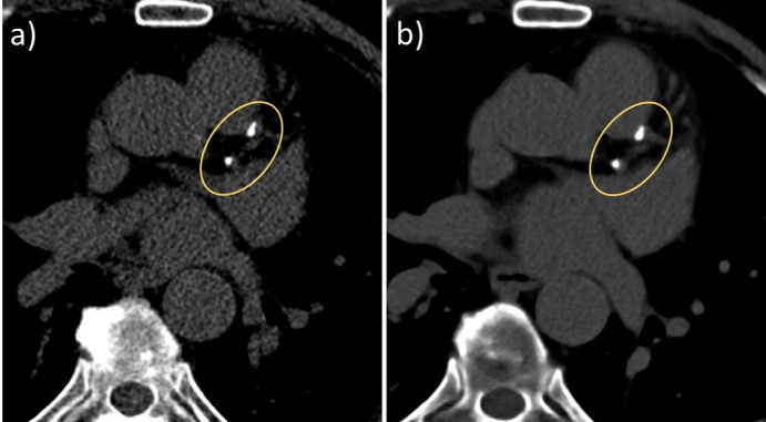
Figure 2.

Clinical example of CAC scoring with PCD-CT. (a) Non-contrast CAC scoring scan of a 73-year-old male patient (120 kVp, 70 keV, 3 mm slice, Qr36, 61 bpm). Agatston score 322. (b) VNI image from a contrast-enhanced scan (120 kVp, 70 keV, 3 mm slice, Qr36, 57 bpm). Agatston score 330. Note the reduced image noise in the VNI image because of the higher radiation dose of the contrast-enhanced scan. CAC, coronary artery calcium; PCD-CT, photon-counting detector CT; VNI, virtual non-iodine.
