. 2023 Oct 25;14(44):12484–12497. doi: 10.1039/d3sc02782a

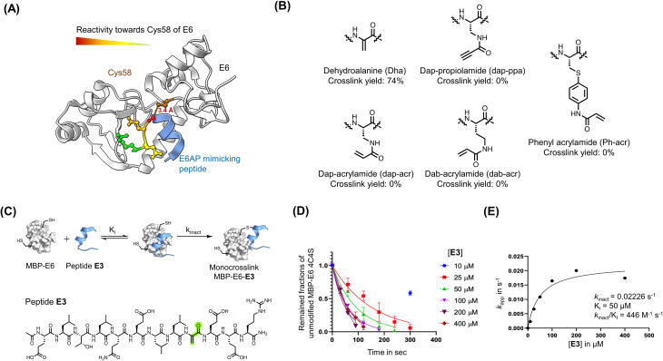
Fig. 2. Dha was installed to generate reactive peptides. (A) Analysis of the main binding interface of 16E6 and E6AP. Cys58 of 16E6 is highlighted in brown. Residues of E6AP were colored based on their measured reactivity towards Cys58 of 16E6: Gly9 in red, Glu10 in orange, Glu11 in yellow, and Arg12 in Green. (B) Structures of electrophiles. (C) A scheme of the bind-and-react strategy. Structure of peptide E3 containing a Dha electrophile highlighted in green. (D) Apparent kinetic constant Kapp was calculated from a kinetic crosslinking study, where 50 nM of MBP–16E6 was incubated with different concentrations of E3. (E) A series of Kapp values were plotted against the corresponding E3 concentration to estimate kinact and Ki.

Fig. 2