
Figure 1.
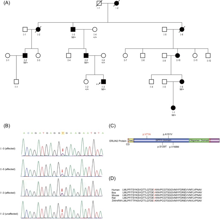
Pedigree of affected family, ERLILN2 variants, and conservation among species. (A) Family pedigree. Black filled symbol, affected; white symbol, unaffected; and black arrows, proband. Sanger sequencing was performed in some subjects (+/+: normal, M/+: heterozygous), demonstrating complete segregation of the ERLIN2 missense variant c.212 T>C, p.V71A with the disease. (B) The partial nucleotide sequences of exon 7 of ERLIN2 show the c.212 T>C variant in the affected or unaffected family members (II‐3, III‐5, IV‐3, and IV‐2). (C) Schematic representation of the basic structure and domain organization of the ERLIN2 protein. The figure was generated using the Illustrator for Biological Sciences (IBS). The observed variants were labeled. The site of the novel variant p.V71A identified in this study is indicated by red color. (D) Sequence alignment of ERLIN2 proteins from various species. The arrows indicate the amino acid changes in this study. Emboldened amino acids are conserved.