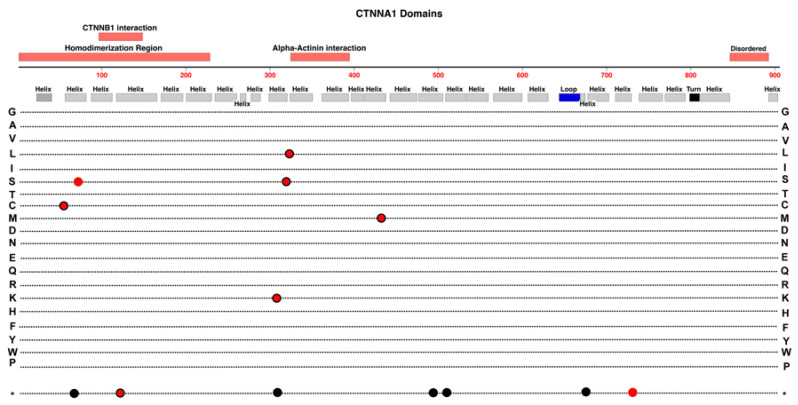
Figure 11.
Structure and functional domain map of variants in CTNNA1 (Catenin alpha-1). Secondary structures are derived from Alphafold data (https://alphafold.ebi.ac.uk/entry/P35221 (accessed on 27 October 2023).) [68,69]. Pathogenic and likely pathogenic variants are indicated for those linked to FEVR (red circles), patterned macular dystrophy (red circles with black outlines), and those linked to non-retinal conditions including cancers (black circles). The bottom row * indicates the location of nonsense (stop codon) variants. Exploration of additional non-pathogenic variants can be found in the UniProt database: https://www.uniprot.org/uniprotkb/P35221 (accessed on 27 October 2023).

