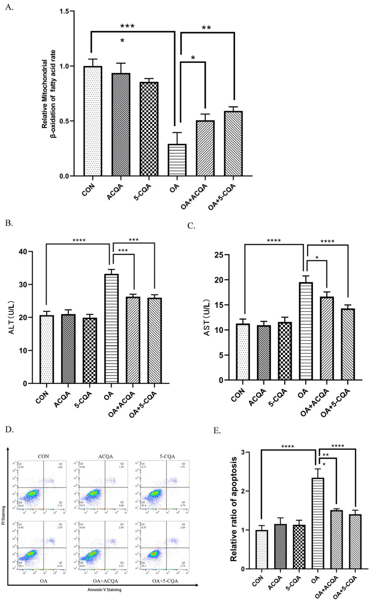
Figure 4.
ACQA enhanced fatty acid β-oxidation and reduced injury caused by steatosis in HepG2 cells. (A) The rate of fatty acid β-oxidation in HepG2 cells was detected. The reduction rate of micromole ferricyanide was used to characterize the β-oxidation rate of fatty acids. (B,C) After the group was treated with chlorogenic acid, the culture medium was collected, and the ALT and AST levels in the culture medium were detected. (D) Apoptosis was assessed with annexin V/PI staining. (E) The data for analysis are expressed as the ratio of total apoptotic cells to the number of normal cells. Data are expressed as mean ± SD, * p < 0.05, ** p < 0.01, *** p < 0.001 and **** p < 0.0001, and compared with OA group, n = 3.

