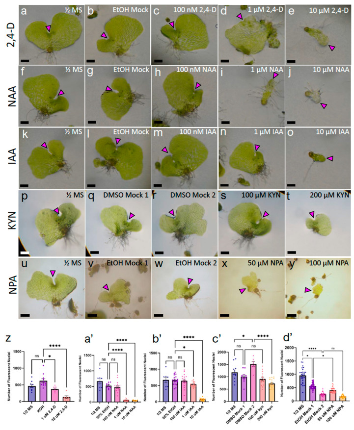
Figure 4.
Hermaphrodites treated with auxin chemicals exhibit decreases in cell proliferation and morphological defects. (a–e,z) An 11-day dosage with 2,4-dichlorophenoxyacetic acid (2,4-D) on gametophyte growth. (f–j,a’) An 11-day dosage with 1-napthaleneacetic acid (NAA). (k–o,b’) An 11-day dosage with indole-3-acetic acid (IAA). (p–t,c’) A 12-day dosage with L-kynurenine (KYN). (u–y,d’) A 14-day dosage with naphthylphthalamic acid (NPA). (a,f,k,p,u) Basal media controls. Solvent mock representatives: (b) 95% EtOH mock for 2,4-D, (g) 95% EtOH mock for NAA, (l) 95% EtOH mock for IAA, (q,r) DMSO mocks for 100 μM and 200 μM KYN, respectively, (v,w) 95% EtOH mocks for 50 μM and 100 μM NPA, respectively. 2,4-D dosage of (c) 100 nM, (d) 1 μM, and (e) 10 μM. NAA dosage of (h) 100 nM, (i) 1 μM, and (j) 10 μM. IAA dosage of (m) 100 nM, (n) 1 μM, and (o) 10 μM. KYN dosage of (s) 100 μM and (t) 200 μM. NPA dosage of (x) 50 μM and (y) 100 μM. (z–d’). Quantification of cell number from individuals grown on auxin chemicals, via DNA staining with Hoechst 33,342 (n ≥ 6, mean ± SEM, (z,d’) Kruskal–Wallis with Dunn’s post-hoc test, (a’,c’) one-way ANOVA with Sidak’s post-hoc test, or (b’) Dunnett’s post-hoc test; ns, not significant; *, p ≤ 0.05; ****, p ≤ 0.0001). Arrowhead pointing at marginal meristem, scale bars = 0.25 mm.

