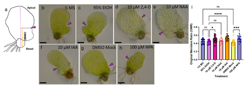
Figure 5.
Exposure to high doses of NPA and IAA during marginal meristem formation alters hermaphrodite meristem location. (a) Diagram of original marginal meristem distance (OMMD) measurement scheme. Long-dashed line marks the apical–basal axis of the prothallus. Solid blue line marks the location of original meristem (magenta arrowhead), perpendicular to the apical–basal line. Orange bracket marks the OMMD. (b–h) Bright field micrographs of live 6-dpp hermaphrodites on treatment media: (b) Basal ½ MS media control. (c) 95% EtOH solvent mock for 2,4-D, NAA, and IAA, (d) 10 μM 2,4-D, (e) 10 μM NAA, (f) 10 μM IAA, (g) DMSO solvent mock for NPA, (h) 100 μM NPA. (i) OMR for treatments shown in (b–h) (n ≥ 14, mean ± SEM, Kruskal–Wallis with Dunn’s multiple comparisons test; ns, not significant; *, p ≤ 0.05; ***, p ≤ 0.001 ****, p ≤ 0.0001). Scale bars = 0.1 mm.

