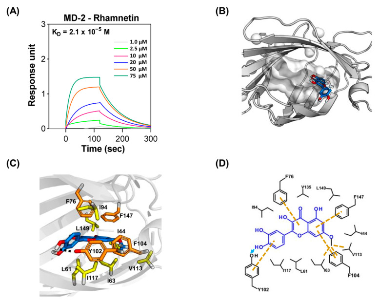
Figure 4.
Binding interactions between MD-2 and rhamnetin. (A) Surface plasmon resonance (SPR) sensorgram of MD-2 interacting with rhamnetin. (B) Overview of the MD-2-rhamnetin complex. The hydrophobic cavity of MD-2 is depicted by the gray surface, and rhamnetin is depicted by the blue stick with red stick for oxygen atoms. (C) Overall complex of the MD-2–rhamnetin. Rhamnetin is depicted by the blue stick with red stick for oxygen atoms, hydrophobic interaction residues are shown as yellow sticks, and important aromatic residues are depicted by orange sticks in MD-2 (gray ribbon cartoon). (D) Two-dimensional diagram of the MD-2–rhamnetin docking pose showing hydrophobic interactions (yellow dash lines) and hydrogen bonding interaction (sky blue arrow).

