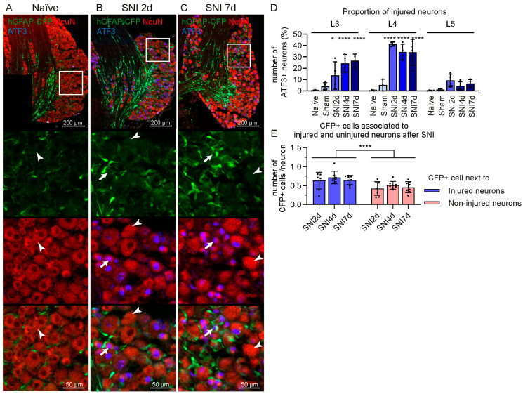
Figure 3.
CFP+ cells are preferentially located in proximity of ATF3+ injured neurons in the DRG after SNI in hGFAP-CFP mice. Representative images of immunofluorescence staining against neuronal marker NeuN (red) and injured neurons marker ATF3 (blue) of L4 DRG from naïve mice (A), at 2 (B) and 7 days after SNI (C). Top panels show the anatomical structure of the DRG, followed by zoomed image (white box) of the endogenous CFP signal (green), antibodies staining (red and blue) and merging, with arrows pointing to NeuN+ ATF3+ injured neurons and arrowheads pointing to NeuN+ ATF3− uninjured neurons. (D) Quantification of ATF3+ neurons in the L3, L4 and L5 DRG at 2, 4 and 7 days after SNI, 7 days after sham surgery, and naïve mice (N = 4 mice, two-way ANOVA: interaction F(8,43) = 8.411 p < 0.0001, post-hoc Dunnett’s multiple comparisons with naïve group). (E) Quantification of CFP+ cells in proximity of ATF3+ and/or ATF3− neurons in the L3 and L4 DRG at 2, 4 and 7 days after SNI. (N = 8 DRGs, two-way ANOVA: adjacent neuron type F(1, 42) = 19.17 p < 0.0001). * p < 0.05, **** p < 0.0001.

