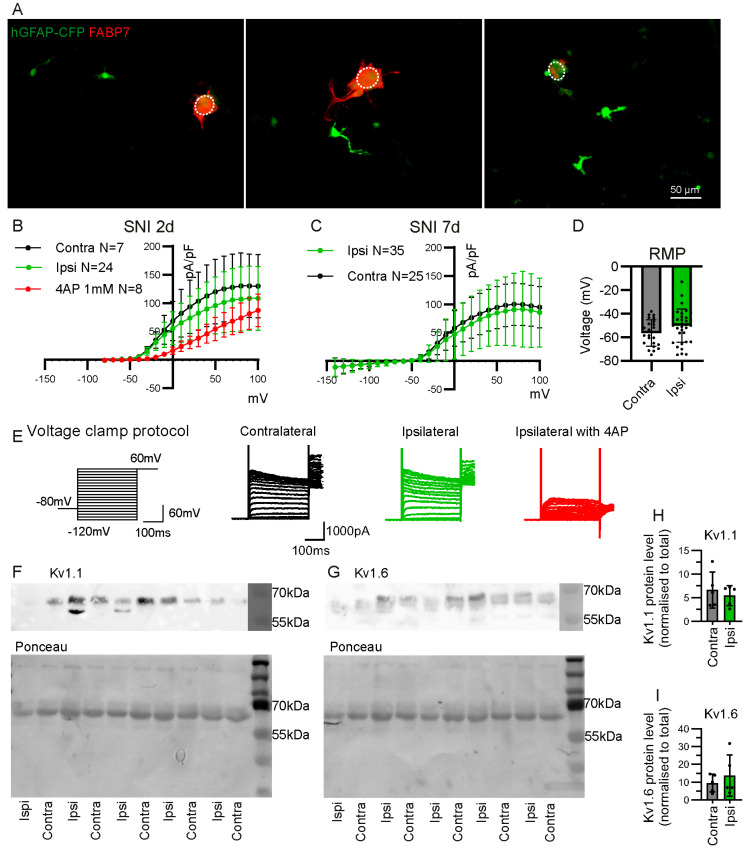
Figure 6.
Electrophysiological characteristics of CFP+ FABP7− cells from the DRG of hGFAP-CFP mice. (A) Representative images of dissociated DRG culture from hGFAP-CFP (green) mice at 7 days after SNI, with immunofluorescence staining against SGC marker FABP7 (red). Dotted circle delineates the neuronal soma. All observed CFP+ cells that are detached from neuronal soma are FABP7−. Potassium current density of detached CFP+ cells in dissociated culture of contralateral or ipsilateral L3, L4 and L5 DRG, at 2 days (B) and 7 days (C) after SNI. The 4AP at 1 mM was used to block Kv currents (two-way ANOVA: B. Interaction Group F(2, 722) = 48.37 p < 0.0001 with post-hoc Dunnett’s multiple comparisons with ipsilateral group ipsi vs. ipsi with 4AP 0 mV to 80 mV p < 0.05, (C) Interaction Group F(1, 1450) = 8.568 p < 0.01 with post-hoc Sidak multiple comparisons contra vs. ipsi −140 mV to 100 mV p > 0.05). (D) Resting membrane potential (RMP) of the patched detached CFP+ cells at 7 days after SNI (contra N = 25 cells, ipsi N = 35 cells, unpaired Student’s t-test p > 0.05). (E) Voltage clamp protocol and representative traces recorded in whole-cell patch clamp from contralateral and ipsilateral cells with and without 4AP application. Western blot for Kv1.1 (F), Kv1.6 (G), with respective ponceau staining for total protein in samples of CFP+ cells sorted by FACS from L3, L4, L5 ipsilateral and contralateral DRG, 7 days after SNI. Quantification of Kv1.1 (H) and Kv1.6 (I) protein level normalised to total protein level (N = 5 samples, paired Student’s t-test p > 0.05).

